Nikon Europe B.V. | Europe & Africa
- pt Change Region
- Global Site
Centro de Excelência
Located in the Lunenfeld-Tanenbaum Research Institute of Sinai Health, Canada’s first Nikon Centre of Excellence seeks to provide access to the latest in imaging technologies to its trainees, visiting scholars, and collaborators at the University of Toronto, the greater Toronto community and around the world. Our goal is to harness the power of advanced imaging and image analysis to address the most salient questions in cell biology and to foster meaningful relationships between our researchers and Nikon’s hardware and software development teams.
Laurence Pelletier, Ph.D.
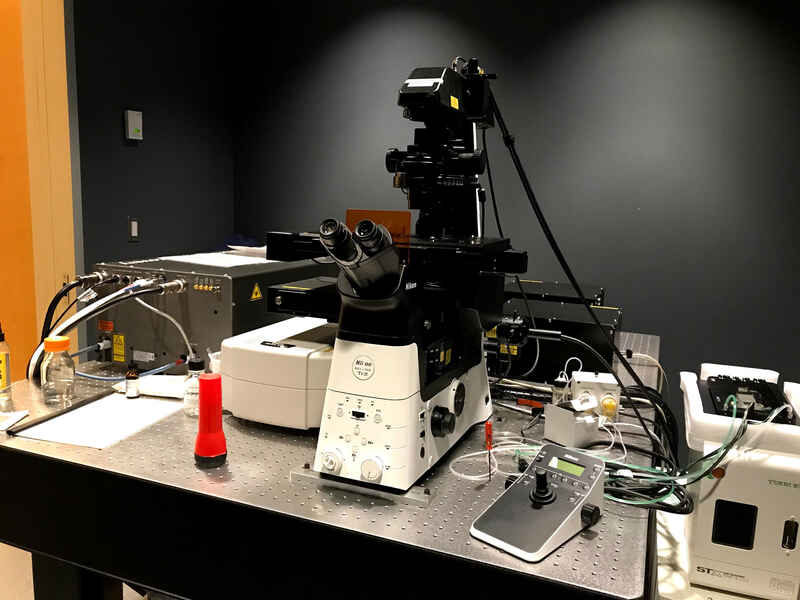
This system features the A1R MP resonant scanning multiphoton system with visible light confocal capabilities, based on the Ti2-E inverted microscope with stage-top incubation chamber, for a variety of live and fixed specimen imaging applications.
NIS-Elements High Content image acquisition and analysis software
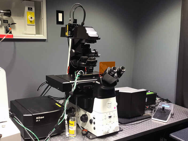
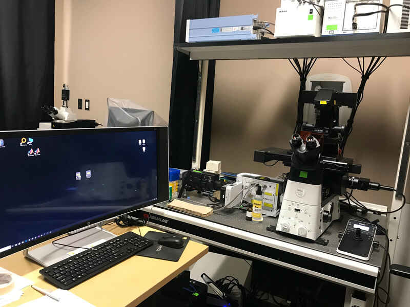
The A1R-HD25 large field of view resonant scanning confocal system and CREST X-LIGHT V2 large field of view spinning disk confocal system are both configured on a Ti2-E inverted microscope, equipped with the stage-top incubation chamber for live cell imaging.
NIS-Elements High Content image acquisition and analysis software
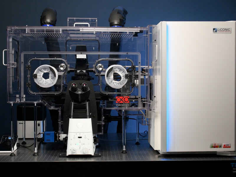
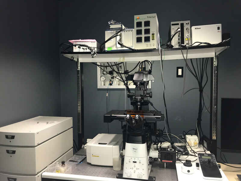
The A1R-HD25 large field of view resonant scanning confocal system on a Ti2-E inverted microscope base, equipped with the full enclosure incubation and robotic delivery system for multiwell plates, to enable long-term high content imaging of live samples.
NIS-Elements High Content image acquisition and analysis software
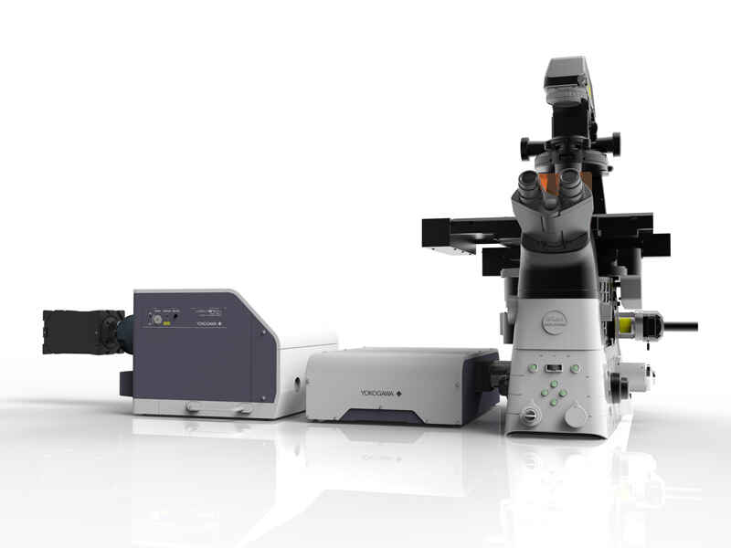
The CSU-W1 SoRa spinning disk with Cairn Research TwinCam optics for simultaneous dual camera imaging, configured on a Ti2-E inverted microscope base, combines confocal and super-resolution capabilities with wide variety of laser lines for fast and gentle multiwavelength imaging.
NIS-Elements High Content image acquisition and analysis software
The CSU-X1 spinning disk confocal system with W-View Gemini emission splitting optics, configured on a Ti2-E inverted microscope base, enables fast and gentle imaging of live and fixed specimens.
NIS-Elements High Content image acquisition and analysis software
The Nikon A1R-HD resonant scanning confocal system with spectral detection capabilities and equipment for high-speed widefield epifluorescence imaging is integrated on a Ti2-E inverted microscope base, for fast multichannel image acquisition.
NIS-Elements High Content image acquisition and analysis software
