- pt Change Region
- Global Site
- Início
- Nikon BioImaging Centers
- Fondazione Istituto Italiano di Tecnologia

Nikon Imaging Center
Fondazione Istituto Italiano di Tecnologia
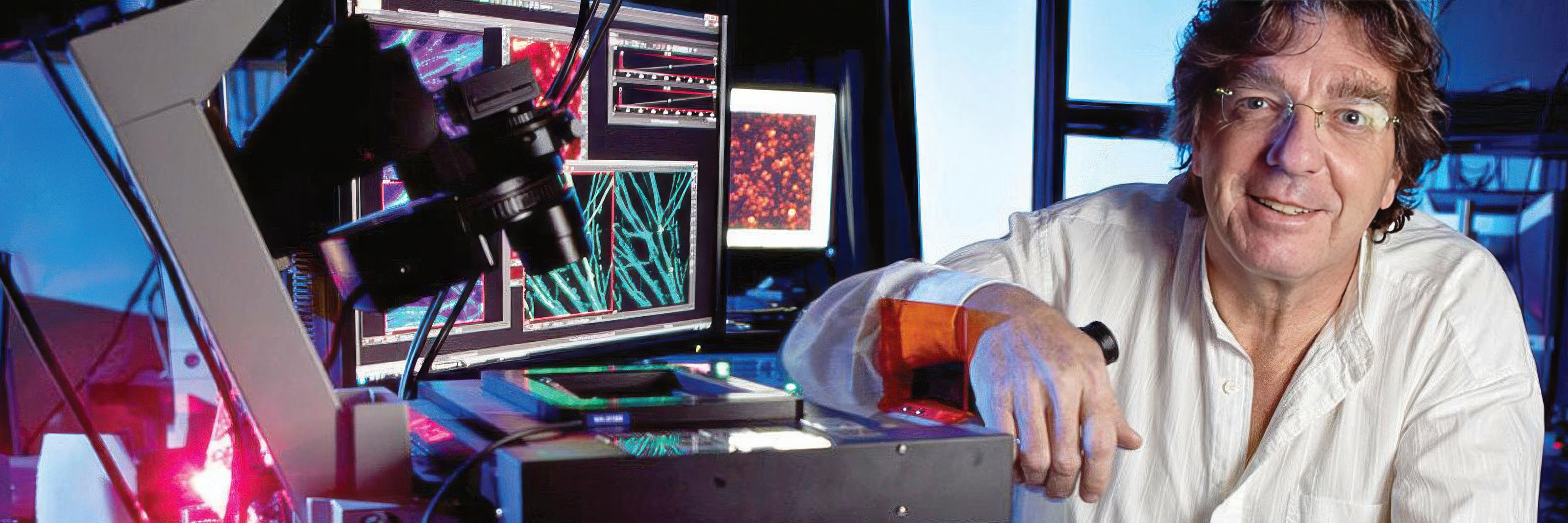
The Nikon Imaging Centre at Fondazione Istituto Italiano di Tecnologia (NIC@IIT) is a core facility for light microscopy developed in partnership between Fondazione Istituto Italiano di Tecnologia and Nikon. The NIC@IIT is to provide to a wide community of scientists and professionals throughout Italy, Europe and the Rest of the World, with the support of Nikon, a large number of up-to-date imaging methodologies to monitor the living cell activity at high spatial and temporal rate. The main expertise of the NIC@IIT is related to Super resolution and multiphoton microscopy, and it is developed in the unique multidisciplinary environment of IIT. In the era of incredible advances in optical microscopy we can state that a new paradigm was born.
The mission of NIC@IIT is to:
- Stimulate innovation in biological research by providing investigators access to cutting edge microscopy resources with a particular emphasis on developing novel imaging solutions to systems biology challenges.
- Support research while giving the researchers access to most advanced technologies in the field of cellular imaging, data analysis and image processing.
- Serve as a training platform providing courses, individual instruction, and consulting on advanced light microscopy techniques.
- Promote cross-discipline collaborations by providing an exciting intellectual commons centered on light microscopy.
- Develop new microscope setups and imaging techniques in response to feedback from the users of NIC@IIT.
- To become a demonstration platform for Nikon’s commercial prospecting.
The NIC@IIT is supported by the Chamber of Commerce of Genoa and by a selected number of partners.
Contato
NIC Director
email hidden; JavaScript is required
Address
Nikon Imaging Centerat Fondazione Istituto Italiano di Tecnologia
Via Morego 30
16163 Genova
Italy
Systems Available
ECLIPSE Ti2-E Inverted Microscope with N-STORM and N-SIM S Super-Resolution
The ECLIPSE Ti2-E / N-SIM S super-resolution microscope is a unique high-speed structured illumination system that achieves acquisition speeds of up to 15 fps, enabling fast biological processes to be captured at twice the spatial resolution of conventional light microscopes (down to 115 nm in XY). The N-STORM super-resolution module uses “Stochastic Optical Reconstruction Microscopy” technology to provide dramatically enhanced resolution that is 10 times that of conventional optical microscopes. Both modalities are combined on the same Ti2-E microscope built for stability with Nikon’s Perfect Focus System (PFS) and Nikon's powerful acquisition and analysis software, NIS-Elements, enabling scientists to achieve true multimodal super-resolution imaging across different scales.
ECLIPSE Ti-E PFS Inverted Microscope System
The ECLIPSE Ti-E with Perfect Focus is Nikon's market leading inverted microscope system offering improved system speed, increased flexibility and efficient multi-mode microscopy as part of a fully-integrated microscope system that is ideal for high-end research and live cell imaging. Perfect Focus is Nikon's exclusive focus correction system that combats axial focus fluctuations in real time during long-term imaging investigations.
A1R MP+ Multiphoton Confocal Microscope
Nikon’s A1R MP+ multiphoton confocal microscope is a unique multiphoton imaging system featuring a fast, high resolution galvanometer scanner and an ultra-high speed resonant scanner that is capable of frame rates from 30 fps at 512 x 512 pixels to as fast as 420 fps in band scan mode. This is especially important in multiphoton microscopy because of the overlap of emission spectra of probes and autofluorescence, which is often unavoidable when using a single laser line.
A1R Confocal Microscope System
The A1R features a "hybrid scanner" (two scanners in one scanning-head). A galvano scanner for low-noise images ranging in resolution from 64 x 64 up to 4096 x 4096 pixels and a resonant scanner that is capable of taking up to 30 full 512 x 512 pixel frames per second. Both scanners can be used in parallel: while the fast resonant scanner is acquiring images, the galvano scanner may perform bleaching or activation of user-defined regions of interest.
- Início
- Nikon BioImaging Centers
- Fondazione Istituto Italiano di Tecnologia
