Nikon Instruments Inc. | Americas
- fr Change Region
- Global Site
Testing for gout and pseudogout is performed by extracting synovial fluid from the affected joint, preparing a wet mount on a microscope slide, and identifying the crystals using polarized light microscopy. The intrinsic optical properties of these crystals make polarized light observation effective for their identification.
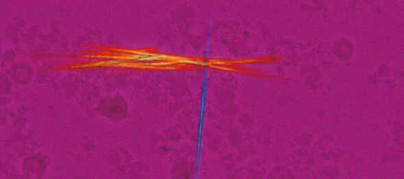
Sodium urate crystals, Sensitive color polarizing, CFI Plan Fluor 40X
Department of Clinical Laboratory, Nihon University Itabashi Hospital
The ECLIPSE Ci-L upright microscope is the most efficient choice offered by Nikon for routine gout testing and other clinical research applications requiring simple or color-sensitive polarized light observation, such as identification of collagen and amyloid. To streamline operation, the Ci-L is available with a combined polarizer and first order red compensator. The polarizing filter and compensator can be independently rotated. Rotation of the compensator allows for simple determination of the sign of birefringence. The ECLIPSE Ni-U upright microscope provides similar capabilities.
The ECLIPSE Ci-POL polarized light microscope is the only Nikon microscope discussed here that is capable of quantitative polarized light microscopy, which allows for precise measurement of birefringence. This may be useful for more in-depth analysis of gout and other crystals in biomedical research settings.
●: Inclus, ⚬: Optionnel
| ECLIPSE Ci-L Upright Microscope |
ECLIPSE Ni-U Upright Microscope |
ECLIPSE Ci-POL Polarized Light Microscope |
|
|---|---|---|---|
| Simple Polarized Light | yes | yes | yes |
| Color-Sensitive Polarized Light | yes | yes | yes |
| Quantitative Polarized Light | no | no | yes |
| Illumination Type | LED | LED or Halogen | LED |
| Motorization | no | Partial available | no |
Gout and pseudogout crystals exhibit the property of birefringence (double refraction), whereby the refractive index (RI) differs between crystal axes due to the molecular architecture. Light incident on a birefringent uniaxial crystal and traveling in a different direction than the crystal optic axis will see two different RIs. Approximately half of the light will be polarized in a direction orthogonal to the optic axis of the crystal and referred to as the “ordinary wave.” The ordinary wave experiences refraction as usual, its path continuing un-deviated through the crystal when incident orthogonal to its surface. The other half of the light is polarized in the same plane as the optic axis and its direction will deviate from that expected due to refraction alone – the “extraordinary wave.”
As the ordinary and extraordinary waves experience different optical path lengths, they become out of phase, resulting in interference colors when recombined into the same plane of polarization. The sign of birefringence is positive when the RI experienced by the extraordinary wave is higher than that of the ordinary wave, and negative under opposite conditions.
Gout crystals are generally long and needle-like while pseudogout crystals are more rhomboid. Gout crystals are negatively birefringent and their long axis is the “fast” axis (corresponding to the extraordinary wave in this case) – light polarized in a plane parallel to the fast axis will pass through the crystal faster than light perpendicular to it because it experiences a lower RI. Pseudogout crystals are positively birefringent, their long axis is the “slow” axis instead.
A polarized light microscope configured for gout testing features a linear polarizer oriented at 0 degrees (east-west) following the light source. When the long (fast) and short (slow) axes of the gout crystal are oriented at ±45 degrees, the energy of the incident light is split equally between these directions. A phase shift results as these two rays traverse the crystal due to the light taking longer to travel via the slow axis.
Later, a second linear polarizing filter – the analyzer – oriented at 90 degrees (north-south) forces the light from these two rays back into the same plane of polarization. The difference in phase becomes manifest as an interference color. Correlating interference color with phase difference and sample thickness can be performed using a Michel-Lévy color chart (Figure 1).
The problem is that small phase differences only result in black-white interference colors. For this reason, a first-order red compensator is used to introduce a 530 nm phase difference (corresponding to a red/magenta tone in the chart). The compensator has its own perpendicular fast and slow axes.
When the long (fast) axis of a gout crystal is aligned with the slow axis of the compensator, the resulting phase difference subtracts from 530 nm, resulting in a yellow-hued interference color (“first order yellow”). However, when the short (slow) axis of a gout crystal is aligned with the slow axis of the compensator, then the phase difference will be greater than 530 nm, resulting in a blue color (“second order blue”). This is illustrated by Figure 2.
Unaffected background light will exhibit a red/magenta color corresponding to the 530 nm phase difference introduced by the compensator. The compensator thus allows the determination of the sign of birefringence using color as a readout, without it both axes would appear a similar shade of gray.
For more information about polarized light microscopy, including theory and microscope configuration, please refer to our MicroscopyU educational article Polarized Light Microscopy.


