Nikon ®
Nikon Healthcare: A Century of Microscopy
Nikon Instruments Inc. | Americas
  • fr Change Region
  • Global Site
  • Artboard 1

Select Your Region and Language

Products and Promotions may differ based on your selected Region.

Americas

  • en English
  • es Español
  • fr Français
  • pr Português

Europe & Africa

  • en English
  • de Deutsch
  • es Español
  • fr Français
  • it Italiano
  • pr Português

Japan

  • ja 日本語

Korea

  • ko 한글

Chinese Mainland

  • zh 简体中文

Asia, Oceania & Middle East

  • en English

Return to the top of the page

  • Produits
  • CRO
  • Applications
  • Ressources
  • BioImaging Centers
  • Service
  • Contactez

Produits de microscope

Light Microscopes

  • Microscopes inversés
  • Microscopes droits
  • Microscopes polarisants
  • Stéréomicroscopes

Advanced Imaging Systems

  • Microscopes confocaux et multiphotoniques
  • Microscopes à super-résolution
  • Criblage cellulaire
  • Photostimulation & TIRF

Microscopes Digitaux

Logiciel

Appareils photos

Optiques

Accessoires

OEM

Produits discontinués

Nikon BioImaging Labs

Nikon BioImaging Labs provide contract research services for microscope-based imaging and analysis to the biotech, pharma, and larger research communities. Each lab's full-service capabilities include access to cutting-edge microscopy instrumentation and software, but also the services of expert biologists and microscopists, who are available to provide quality cell culture, sample preparation, data acquisition, and data analysis services.

Boston, United States

Europe

Shonan, Japan

Applications

Sciences de la vie

  • Deep Learning en microscopie
  • Découverte de médicament
  • Électrophysiologie
  • Imagerie à haut débit
  • Imagerie de cellules vivantes
  • Microinjection
  • Imagerie à l'échelle nanométrique
  • Imagerie organoïde et organe sur puce
  • Médecine régénérative
  • Culture tissulaire
  • Imagerie de l'organisme entier et des tissus

Recherche clinique

  • Assisted Reproductive Technology (ART)
  • Gout Testing
  • Imaging Fixed Slides
  • Slide Scanning

Éducation

OEM

Ressources

Featured Resources

  • PubScope

    Search and filter over 125,000 Open Access Articles that utilize Nikon products and supported third party systems.

  • Téléchargements de logiciels/micrologiciels

    Téléchargez des programmes de logiciels et de micrologicielss pour les produits de microscope Nikon.

  • Guides en ligne

    Guides étape par étape pour l'alignement et l'utilisation de certains microscopes Nikon.

  • Essai gratuit de Denoise.ai

    Denoise.ai supprime automatiquement le bruit de Poisson des images confocales - essayez-le gratuitement.

  • MicroscopyU

    MicroscopyU de Nikon est la principale source d'informations pédagogiques sur la microscopie optique.

  • Sélecteur d’objectifs

    Trouvez l'objectif Nikon adapté à votre flux de travail.

  • Nikon Small World

    Le site Web Nikon Small World - tout sur les concours d'images/vidéos au microscope.

Browse Resources

Service

À propos

  • Nouvelles
  • Profil de la société
  • Carrières
  • Durabilité
  • Accueil
  • CRO
  • Europe
  • Applications
  • Histological Image Segmentation

Nikon BioImaging Labratories

Europe

  • Boston, United States
  • Shonan, Japan
  • Vue d’ensemble
  • Applications
  • Research Services
  • Expertise
  • FAQs
  • Resources
  • News & Events

Histological Image Segmentation

Histological image analysis is a crucial aspect of medical research and diagnostics. It involves the microscopic examination of tissue samples that have been stained to highlight different cellular components and structures. This analysis allows pathologists to diagnose diseases, understand cellular interactions, and conduct detailed anatomical studies. The accuracy of histological analysis is vital, as it directly impacts clinical decisions and the understanding of complex biological processes.

Human cervical tissue with H&E staining
Tumor mass segmentation
Red blood cells segmentation

Challenges

The intricacies of histological image segmentation stem from the way these images are typically obtained. In histology, the stains and dyes applied to tissue samples are absorptive rather than fluorescent. They are visualized in full color under white light illumination, and unlike in fluorescence microscopy, the data from various stains are not distinctly separated based on light emission. Thresholding, a simple segmentation method that separates objects based on pixel intensity, often falls short due to the variability in stain absorption and the complex morphology of tissues, including the presence of various cell types with diverse shapes and sizes. Consequently, the segmentation and analysis of histological images have traditionally relied heavily on manual inspection by experienced professionals, a process that is both time-consuming and subjective.

Solutions

Artificial intelligence (AI) offers a solution for the precise and efficient segmentation of histological images. Deep learning-based algorithms can segment images of complex tissue samples with human-like accuracy, learning from annotated examples to identify numerous features swiftly. Once trained, AI can quickly discern thousands of elements in images, streamlining pre-clinical research by reducing manual effort and enhancing the discovery of disease pathways and treatment options.

Method

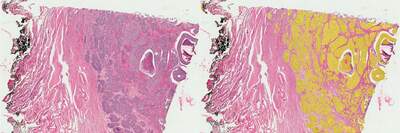
In our study, we utilized AI to enhance the analysis of histological slides, focusing on a human cervical cancer specimen stained with the conventional Hematoxylin and Eosin (H&E) method. This staining technique is intentionally nonspecific—hematoxylin colors nuclei blue and eosin imparts a pink hue to the cytoplasm, thus providing a stark contrast that facilitates the examination of the entire tissue section. Despite its simplicity, the H&E method introduces challenges in segmentation, as it requires careful consideration of the brightness of the stain and the diverse sizes and shapes of cellular structures.

To address these challenges, we developed multiple AI networks, each tailored to identify and differentiate features across varying scales, from large tumor masses down to red blood cells and single cell nuclei. These networks underwent a training process using a small, annotated segment of the histological image and were subsequently deployed to analyze the full extent of the tissue sample.

Results

Our analysis revealed that the tumor regions accounted for approximately 21% of the total tissue area examined. The AI-driven segmentation process successfully delineated around 360,000 nuclei, with 132,000 of those located within the tumor areas.

Conclusion

The application of the trained neural networks allowed for the rapid processing of an extensive 9.5 x 7 mm image in a matter of seconds. These results are promising for future studies, as the networks could be adapted for analyzing other H&E-stained tissues, either in their current form or with minor modifications to accommodate different sample characteristics, thus significantly accelerating the pre-clinical research workflow.

Contactez

  • Accueil
  • CRO
  • Europe
  • Applications
  • Histological Image Segmentation
Nikon ®
  • LinkedInLinkedIn
  • BlueskyBluesky
  • FacebookFacebook
  • InstagramInstagram
  • ThreadsThreads
  • YouTubeYouTube
  • XX

Contact Nikon Instruments Inc.

+1-631-547-8500
+1-800-52-NIKON (within the U.S.A. only)
nikoninstruments.us@nikon.com

À propos

  • Nouvelles
  • Événements
  • Profil de la société
  • Carrières
  • Durabilité
  • Bien-être

Popular Links

  • Dernières nouvelles et actualités
  • Sélecteur d’objectifs
  • Resolution Calculator
  • PubScope
  • OEM
  • Nikon Small World
  • MicroscopyU

Autres Produits Nikon

  • Produits d'imagerie
  • Produits de métrologie industrielle
  • Systèmes de lithographie à semi-conducteurs
  • Systèmes de lithographie à FPD
  • Contactez
  • Plan du site
  • Confidentialité
  • Cookie settings
  • Do Not Sell or Share My Personal Information
  • Conditions d’utilisation
  • Carrières

© 2025 Nikon Instruments Inc.