Nikon Instruments Inc. | Americas
Artboard 1
fr Change Region

Global Site

The Scripps Research Institute

Contactez

Centre d’excellence

The Scripps Research Institute

Researchers at The Scripps Research Institute (TSRI) can now probe more deeply and clearly into the microscopic elements of cells with the recent opening of a new Nikon Center of Excellence on the California campus, featuring the latest in advanced imaging technology.

Contactez

CofE Director

email hidden; JavaScript is required

Address

The Scripps Research Institute
Dept. of Molecular and Cellular Neuroscience
10550 N. Torrey Pines Road
DNC 210
La Jolla, CA 92037


Systems Available

CresOptics X-Light V3 Spinning Disk Confocal

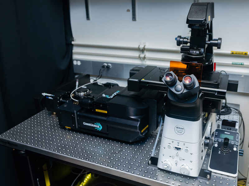
Utilizing a CrestOptics X-Light V3 spinning disk integrated with a Nikon ECLIPSE Ti2 and a Photometrics Kinetix camera, this system is ideal for any experiment where imaging speed and sample health is a priority.

Components

  • Fully motorized ECLIPSE Ti2-E with Perfect Focus System and 4-channel epifluorescence
  • Crest V3 large field of view spinning disk with Lumencor Celesta 7-line solid state laser light source
  • Photometrics Kinetix large field of view 95% QE camera
  • 2X/4X/10X/40X air objectives with DIC
  • High speed Prior Queensgate 600 µm piezo
  • NIS-Elements with high content acquisition tools

Gattaca iLas2 TIRF/Photostimulation + STORM System

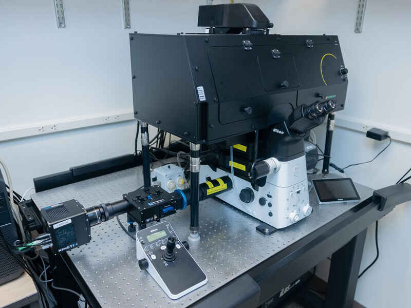
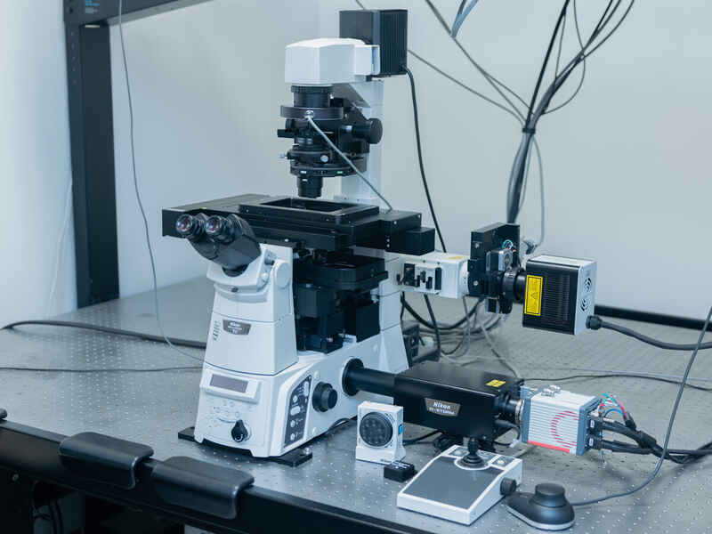
This system combines the iLas2 ring TIRF/FRAP module with capabilities for STORM imaging as well. This system is well suited for imaging smaller structures both axially and laterally.

Components

  • Gattaca iLas2 TIRF/photostimulation illuminator
  • Spectra III 7-line light source
  • LAPP combined illumination module
  • Photometrix Kinetix 10 MP sCMOS
  • Cairn OptoSplit II
  • Polygon1000 dynamic spatial illuminator
  • Tokai Hit incubation chamber
  • PE100-NIF Peltier stage
  • iXON Life 888 back-illuminated EMCCD camera
  • STORM with cylindrical lens
  • CFI60 Apo 40X Lambda S LWD Water immersion objective, with water immersion dispenser

AX R Point Scanning Confocal System

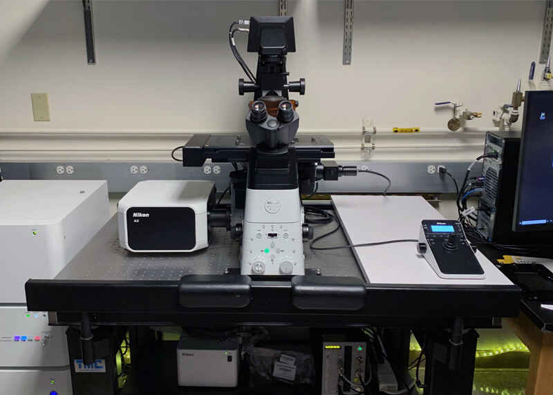
This system is equipped with the highly flexible AX R confocal system, which allows for fast, gentle high-resolution imaging of large tissue samples.

Components

  • 8K galvo / 2K resonant imaging modes
  • ECLIPSE Ti2-E inverted microscope with Perfect Focus System (PFS)
  • LUA-S4 4-line laser unit (405/488/561/640 nm)
  • DUX-VB4 spectral detection
  • NIS-Elements software

A1R Multiphoton Confocal System

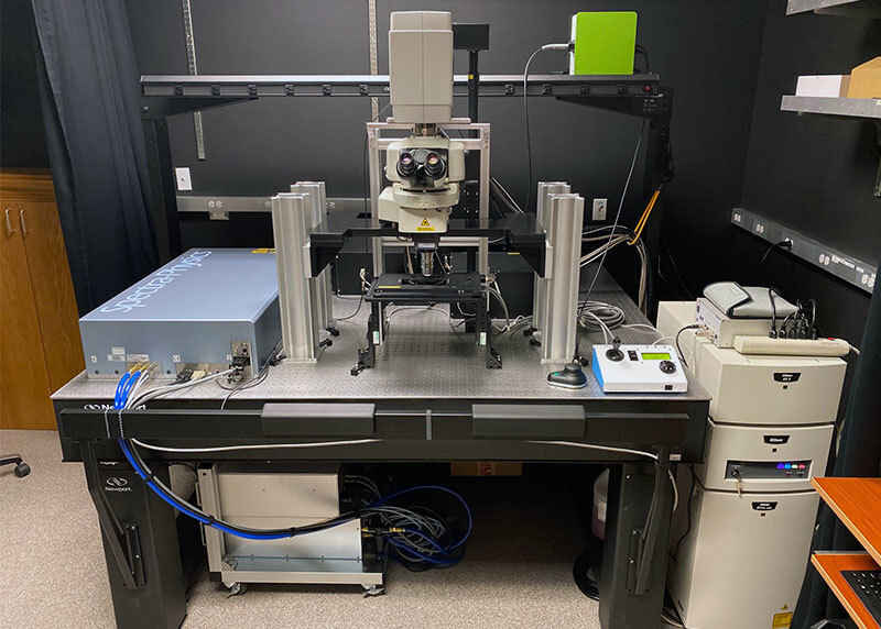
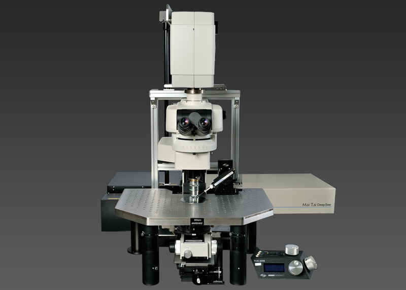
Utilizing an open frame design, this multiphoton system is ideal for deep tissue imaging, with galvo and resonance imaging capabilities and the flexibility to integrate in vivo small animal experimental setups.

Components

  • 4K galvo / 1K resonant imaging modes
  • Open frame upright microscope
  • Removable ASI XYZ motorized stage
  • Spectra-Physics Insight X3 Dual IR 1300 nm Laser
  • LUN4 4-line visible laser unit (405/488/561/640 nm)
  • Non-descanned detector (NDD)
  • DU4G descanned detection
  • NIS-Elements software

ECLIPSE Ti2 Widefield Microscope

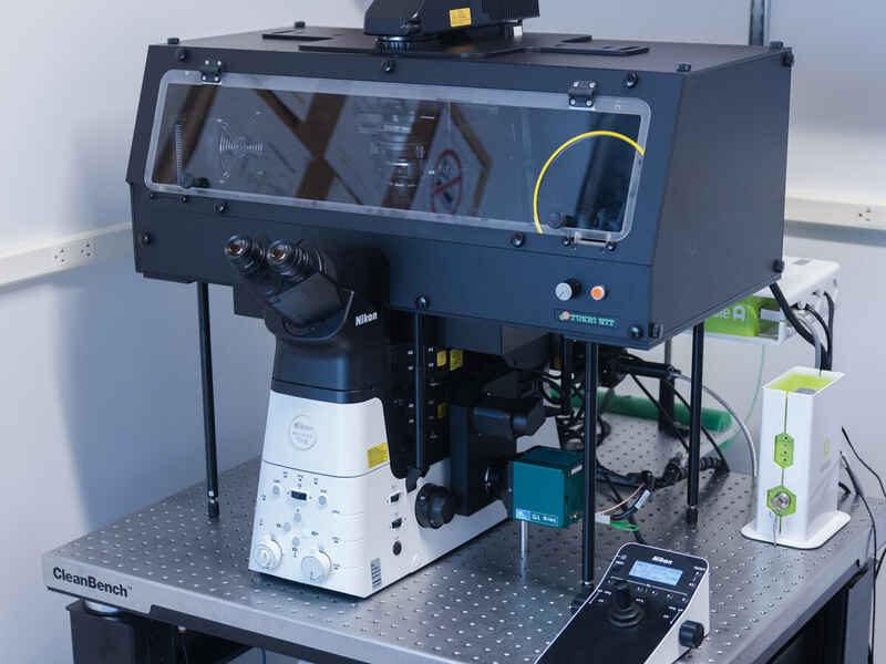
This widefield system is equipped with a Tokai Hit incubation chamber, and is ideal for long term imaging in live cell samples.

Components

  • ECLIPSE Ti2 inverted microscope
  • Photometrics Iris 15 camera
  • Tokai Hit incubation chamber
  • Spectra III light engine
  • Alveole PRIMO micropatterning

N-STORM/TIRF/A1Rsi Super-Resolution & Confocal System

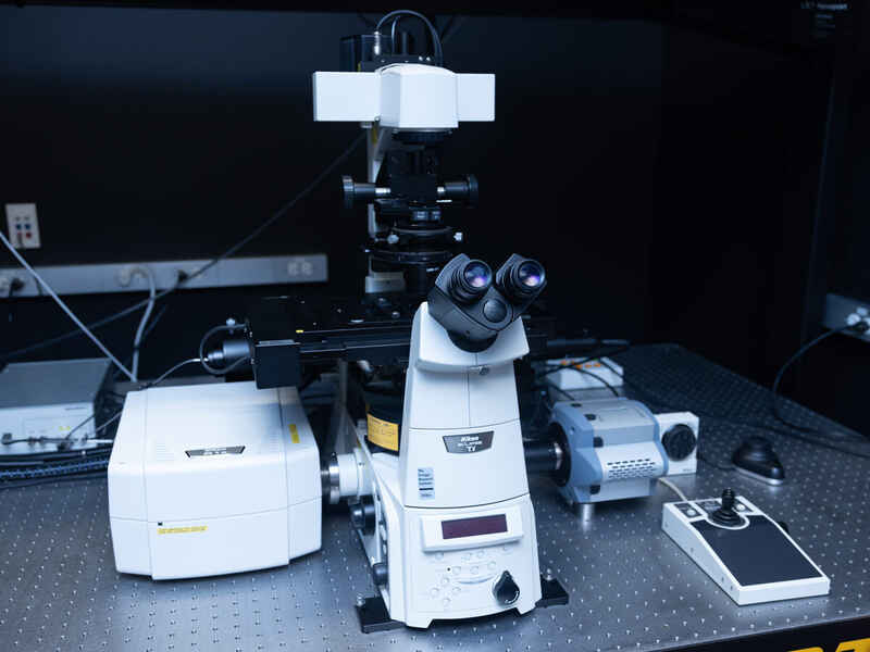
Featuring both the N-STORM single molecule localization and A1R HD resonant scanning confocal, this system is ready for applications ranging from super-resolution to fast live-cell confocal or TIRF.

Components

  • Ti-E inverted microscope with Perfect Focus System (PFS)
  • A1-DUS 32-channel spectral detector
  • N-STORM super-resolution single molecule localization microscopy system
  • LU4A 4-line laser unit (405nm, 488nm, 561nm, 638nm)
  • Platine Z piezo série Mad City Labs Nano-Z
  • DU4 4-channel confocal detector
  • Andor iXon 897 EMCCD camera
  • NIS-Elements software

A1Rsi Resonant Scanning Confocal System

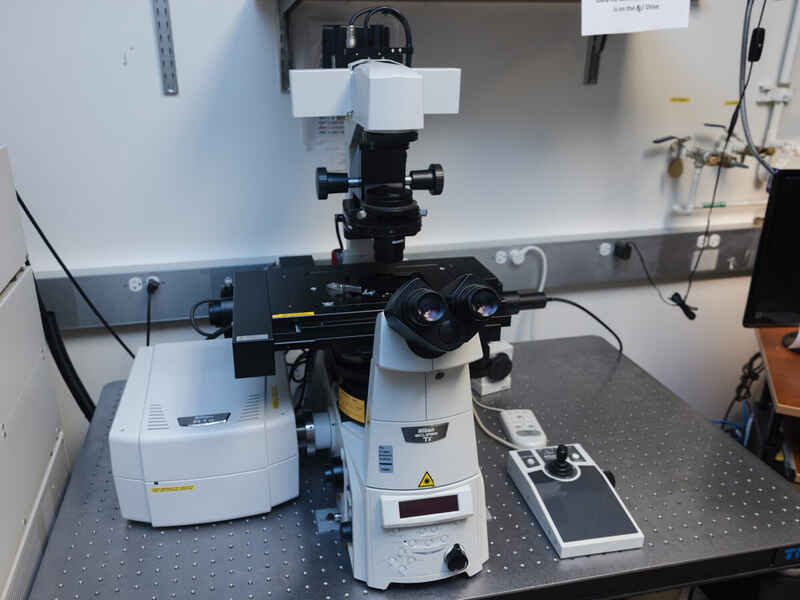
Using the Nikon A1Rsi resonant scanning confocal system with spectral detector, this system is made for linear unmixing of multi-labeled samples.

Components

  • Ti-E inverted microscope with Perfect Focus System (PFS)
  • A1Rsi resonant scanning confocal system
  • LUN-4 4-line laser unit (405nm, 488nm, 561nm, 640nm)
  • A1-DUS 32-channel spectral detector
  • DU4G GaAsP 4-channel confocal detector
  • NIS-Elements software

C2 Point Scanning Confocal System

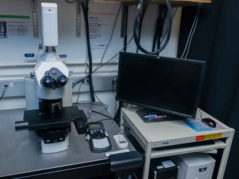
This C2 point-scanning confocal system is configured on a 90i upright microscope and is a great option for applications requiring optical sectioning.

Components

  • C2 point scanning confocal system
  • LU4A 4-line laser unit (405nm, 488nm, 561nm, 638nm)
  • DU3 3-channel confocal detector
  • NIS-Elements software

A1R Resonant Scanning Confocal System (Upright)

This A1R resonant scanning confocal system is configured on an FN1 upright microscope, and providing added flexibility for larger samples.

Components

  • FN1 upright microscope
  • LUN-4 4-line laser unit (405nm, 488nm, 561nm, 640nm)
  • DU4G GaAsP 4-channel confocal detector
  • NIS-Elements software

Système à super-résolution N-STORM/TIRF

While this microscope features the N-STORM localization microscopy system for super-resolution, it can also be operated as a fast live-cell TIRF microscope.

Components

  • Ti-E inverted microscope with Perfect Focus System (PFS)
  • N-STORM super-resolution single molecule localization microscopy system
  • Unité laser à 4 lignes LUN-V (405 nm, 488 nm, 561 nm, 647 nm)
  • Platine Z piezo série Mad City Labs Nano-Z
  • Hamamatsu ORCA-Flash4.0 V2+ sCMOS camera
  • World Precision Instruments AirTherm stage heater
  • In Vivo Scientific CO2 controller
  • NIS-Elements software