


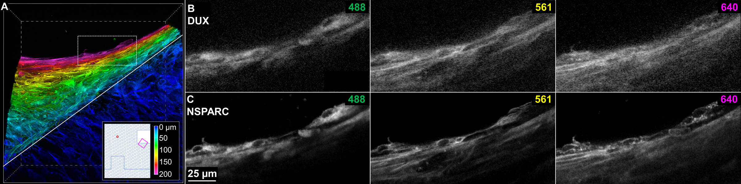
Figure 2: Optimized imaging conditions reveal cellular detail. (A) Depth-coded 3D view of cells proliferating off the edge of the block. The 442 x 442 x 204 μm volume was imaged in resonant scanning mode using the 20x water immersion objective and NSPARC detector. In total, 116, 1024 px frames were collected over 73 seconds. Solid grey line indicates underlying Bio-Block structure. (B, C) Comparison of cells imaged through 200 μm of media using (B) the 20x Plan Apo λD air objective and traditional confocal detector versus (c) the 20x Apo LWD λS water-immersion objective and NSPARC detector (C). This is a composite extended-depth of focus (EDF) image covering 5.28 μm of depth within the white-boxed region in panel A.