Nikon Instruments Inc. | Americas
- pt Change Region
- Global Site
The Nikon Bioimaging Lab (NBIL) can help you advance your research to the next level. Consult with our expert PhD-level staff to plan, execute, and analyze your imaging data, and take advantage of the latest imaging technologies.
We can support your needs throughout the entire imaging pipeline. We can process and embed your tissue, section and create slides for staining, provide antibody optimization services, perform low- and high-plex staining, followed by imaging and analysis our on state-of-the-art systems.
We offer cell culture services for expansion of your cell line for downstream experiments. These services include 2D flask expansion, expansion from a frozen stock, cell banking, and organoid generation.
The Nikon BioImaging Lab is equipped with all necessary accessories and equipment to successfully culture and manipulate your cells or tissues. Once the team grows your cells of interest, we can develop your assay and perform it in-house. NBIL has access to more complex culture models provided by leading biotech organizations and tissue modeling companies.
Both fixed and live cells can be transported to the NBIL. For precious live-cell transport, we make use of CellBox GmbH’s services., ensuring that we can safely transport any number of plates when the need arises.
Together with the Nikon BioImaging Lab, you can now validate your hypotheses, select the optimal performer from a compound library and test for off-target toxicities at higher fidelity and precision than you thought possible.
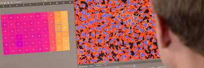
At the Nikon BioImaging Lab, we have access to a wide variety of imaging setups and experts making us comfortable to take on any imaging challenge. We have state-of-the-art confocal microscopes along with the latest software, which can perform automated acquisition in combination with on-the-fly analysis, which includes the use of AI. For complicated analysis questions, our in-house experts can find the answer to any research question.
We can perform varying levels of image analysis and report custom results based on your specific needs. This applies to images taken at NBIL or taken elsewhere. We can also aid with custom solutions for complex questions leveraging our AI/Machine learning software.
You can rely on the Nikon BioImaging Lab to provide accurate reporting to meet your needs. In addition, all services provided are Quality Controlled and ensures that the reporting provided adheres to a defined set of quality criteria as you require.


