Nikon
Nikon Healthcare: A Century of Microscopy
尼康精机(上海)有限公司 | Chinese Mainland
  • zh Change Region
  • Global Site
  • Artboard 1

Select Your Region and Language

Products and Promotions may differ based on your selected Region.

Americas

  • en English
  • es Español
  • fr Français
  • pr Português

Europe & Africa

  • en English
  • de Deutsch
  • es Español
  • fr Français
  • it Italiano
  • pr Português

Japan

  • ja 日本語

Korea

  • ko 한글

Chinese Mainland

  • zh 简体中文

Asia, Oceania & Middle East

  • en English

Return to the top of the page

  • 产品和服务
  • 解决方案
  • 资料
  • 图像中心
  • CRO
  • 关于我们
  • 联系我们

显微镜产品

光学显微镜

  • 倒置显微镜
  • 正置显微镜
  • 偏光显微镜
  • 体视显微镜

高级成像显微系统

  • 共聚焦和多光子显微镜
  • 超分辨显微镜
  • 高内涵成像
  • 光刺激和TIRF

数码显微镜

软件

相机

物镜

附件

OEM

已停产的产品

Nikon BioImaging Labs

Nikon BioImaging Labs provide contract research services for microscope-based imaging and analysis to the biotech, pharma, and larger research communities. Each lab's full-service capabilities include access to cutting-edge microscopy instrumentation and software, but also the services of expert biologists and microscopists, who are available to provide quality cell culture, sample preparation, data acquisition, and data analysis services.

Boston, United States

Europe

Japan

解决方案

生命科学

  • 显微成像中的深度学习
  • 药物研发
  • 电生理学
  • 高通量成像
  • 活细胞成像
  • 显微注射
  • 纳米级成像
  • 类器官和器官芯片成像
  • 再生医学
  • 细胞培养
  • 完整生物体和组织成像

临床研究

  • 痛风检测
  • 固定玻片成像
  • 玻片扫描

教学显微镜

OEM

资料

实用信息

  • PubScope (English)

    Search and filter over 125,000 Open Access Articles that utilize Nikon products and supported third party systems.

  • 软件/固件下载

    下载尼康显微镜产品相关的软件及固件程序。

  • 物镜选择器

    找到最适于您工作流程的尼康物镜。

  • 在线指南

    调整和操作精选尼康显微镜的步骤指南。

  • 网络研讨会

    关于光学显微镜技术及其应用的网络研讨会。

  • MicroscopyU

    通过尼康MicroscopyU网站为您介绍有关光学显微镜的教学资讯。

资料一览

关于我们

  • 最新消息
  • 接下来的活动
  • 可持续发展
  • 主页
  • CRO
  • Boston, USA
  • Applications
  • Whole Slide Scanning

Nikon BioImaging Labratories

Boston, USA

  • Europe
  • Japan
  • 概览
  • Applications
  • Research Services
  • Expertise
  • FAQs
  • Resources
  • News & Events

Whole Slide Scanning

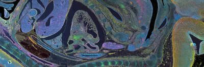
We provide whole slide imaging services, brightfield or fluorescence, of fixed specimens for histology, pathology, gene expression, and spatial biology studies.

Rat cerebellum stained for calbindin (red), NF-L (green), nuclei (blue)
Papanicolaou stain for cervical cancer
HER2 staining for breast cancer
H&E staining for colon cancer

Challenges

Typical commercial digital slide scanners are often low magnification, high NA objectives. At NBIL slides can be imaged similar to commercial digital slide scanners, and desired regions of interests can be further imaged using high magnification, high NA objectives.

Solutions

At Nikon we’re known for our unparalleled image quality. NBIL is equipped with Nikon’s objective lineup, designed to image a variety of samples at different magnifications. In addition, at NBIL you have access to Nikon’s diverse portfolio of imaging modalities from widefield to super resolution based on the requirements of your biological sample and scientific question. At NBIL we leverage Nikon’s NIS Elements software and its AI toolbox to increase experimental throughput and maximize data output. We offer custom AI-trained solutions to address your customer-specific questions, and we deliver your data based on your output requirements.

  • 主页
  • CRO
  • Boston, USA
  • Applications
  • Whole Slide Scanning
Nikon
  • 微信微信

关于我们

  • 可持续发展
  • Well-being

相关网站

  • 物镜选择器
  • PubScope
  • OEM
  • Nikon Small World
  • MicroscopyU

其他尼康产品

  • 映像产品
  • 工业测量产品
  • 半导体光刻系统
  • FPD光刻系统
  • 联系方式
  • 网站地图
  • 隐私
  • 使用条款

© 2025 尼康精机(上海)有限公司