
尼康精机(上海)有限公司 | Chinese Mainland
- zh Change Region
- Global Site
2026年9月
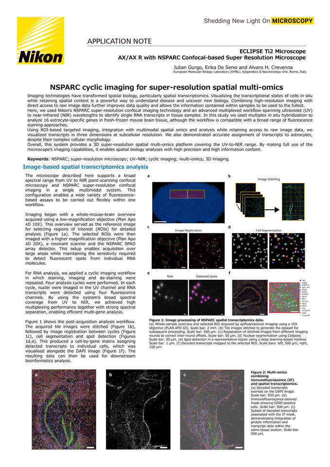
Imaging technologies have transformed spatial biology, particularly spatial transcriptomics. Visualizing the transcriptional states of cells in situ while retaining spatial context is a powerful way to understand disease and uncover new biology. Combining high-resolution imaging with direct access to raw image data further improves data quality and allows the information contained within samples to be used to the fullest.
Here, we used Nikon’s NSPARC super-resolution confocal imaging technology and an advanced multiplexed workflow spanning ultraviolet (UV) to near-infrared (NIR) wavelengths to identify single RNA transcripts in tissue samples. In this study we used multiplex in situ hybridization to analyze 16 astrocyte-specific genes in fresh-frozen mouse brain tissue, although the workflow is compatible with a broad range of fluorescence staining approaches.
Using ROI-based targeted imaging, integration with multimodal spatial omics and analysis while retaining access to raw image data, we visualized transcripts in three dimensions at subcellular resolution. We also demonstrated accurate assignment of transcripts to astrocytes, despite their complex cellular morphology.
Overall, this system provides a 3D super-resolution spatial multi-omics platform covering the UV-to-NIR range. By making full use of the microscope’s imaging capabilities, it enables spatial biology analyses with high precision and high information content.
Keywords: NSPARC; super-resolution microscopy; UV–NIR; cyclic imaging; multi-omics; 3D imaging.