- zh Change Region
- Global Site
应用手册

The mysterious and resilient creature in the moss around us, the tardigrade - Let's catch it! Let's observe it! -
2024年6月
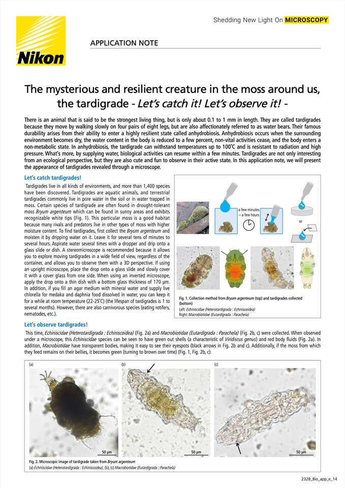
There is an animal that is said to be the strongest living thing, but is only about 0.1 to 1 mm in length. They are called tardigrades because they move by walking slowly on four pairs of eight legs, but are also affectionately referred to as water bears. Their famous durability arises from their ability to enter a highly resilient state called anhydrobiosis. Anhydrobiosis occurs when the surrounding environment becomes dry, the water content in the body is reduced to a few percent, non-vital activities cease, and the body enters a non-metabolic state. In anhydrobiosis, the tardigrade can withstand temperatures up to 100℃ and is resistant to radiation and high pressure. What's more, by supplying water, biological activities can resume within a few minutes. Tardigrades are not only interesting from an ecological perspective, but they are also cute and fun to observe in their active state. In this application note, we will present the appearance of tardigrades revealed through a microscope.
Let's catch tardigrades!
Fig. 1. Collection method from Bryum argenteum (top) and tardigrades collected (bottom)
Left: Echiniscidae (Heterotardigrada : Echiniscoidea)
Right: Macrobiotidae (Eutardigrada : Parachela)
Tardigrades live in all kinds of environments, and more than 1,400 species have been discovered. Tardigrades are aquatic animals, and terrestrial tardigrades commonly live in pore water in the soil or in water trapped in moss. Certain species of tardigrade are often found in drought-tolerant moss Bryum argenteum which can be found in sunny areas and exhibits recognizable white tips (Fig. 1). This particular moss is a good habitat because many rivals and predators live in other types of moss with higher moisture content. To find tardigrades, first collect the Bryum argenteum and moisten it by dripping water on it. Leave it for several tens of minutes to several hours. Aspirate water several times with a dropper and drip onto a glass slide or dish. A stereo microscope is recommended because it allows you to explore moving tardigrades in a wide field of view, regardless of the container, and allows you to observe them with a 3D perspective. If using an upright microscope, place the drop onto a glass slide and slowly cover it with a cover glass from one side. When using an inverted microscope, apply the drop onto a thin dish with a bottom glass thickness of 170 μm. In addition, if you fill an agar medium with mineral water and supply live chlorella for medaka and daphnia food dissolved in water, you can keep it for a while at room temperature (22-25℃) (the lifespan of tardigrades is 1 to several months). However, there are also carnivorous species (eating rotifers, nematodes, etc.).
Let's observe tardigrades!
This time, Echiniscidae (Heterotardigrada : Echiniscoidea) (Fig. 2a) and Macrobiotidae (Eutardigrada: Parachela) (Fig. 2b, c) were collected. When observed under a microscope, this Echiniscidae species can be seen to have green out shells (a characteristic of Viridiscus genus) and red body fluids (Fig. 2a). In addition, Macrobiotidae have transparent bodies, making it easy to see their eyespots (black arrows in Fig. 2b and c). Additionally, if the moss from which they feed remains on their bellies, it becomes green (turning to brown over time) (Fig. 1, Fig. 2b, c).
Fig. 2. Microscopic image of tardigrade taken from Bryum argenteum
(a) Echiniscidae (Heterotardigrada : Echiniscoidea), (b), (c) Macrobiotidae (Eutardigrada : Parachela)
Let's observe the details!
Fig. 3. Tardigrade claws
(a) Macrobiotus shonaicus, (b) claws of Macrobiotus shonaicus, (c) claws of Macrobiotidae, which belong to the same family of Macrobiotus shonaicus, (d) claws of Echiniscidae
Microscope : ECLIPSE Ti2, Camera : Digital Sight 10, Objective lens :CFI Plan Fluor Lambda D 40X
Next, the details were observed using Macrobiotus shonaicus (Tardigrada: Macrobiotidae), kindly provided by Dr. Kazuharu Arakawa (Institute for Advanced Biosciences, Keio University). This species was described in 2018 by Professor Arakawa and his collaborators in Yamagata Prefecture, Japan. The eyespots appear larger (Fig. 3, 4, 5) than the species collected in Kanagawa Prefecture, Japan (Fig. 2b, c).
It also has four distinctive pairs of limbs, each with four claws (Fig. 3a). Of the four pairs of limbs, only the last pair has claws facing backwards, while the rest have claws facing forward (Fig. 3a). The way the claws grow differs greatly between the Macrobiotidae and the Echiniscidae (Fig. 3b, c, d). The length and shape of these claws are one of the key points to distinguish tardigrade species.
Next, the mouth was observed. It looks like a bow and arrow, with a tube extending from its small, round mouth and slightly arched tubes on both sides of the tube (Fig. 4). The round pharynx is located further back (Fig. 4, bottom middle). Tardigrades extend toothed needles from their mouths, trap food, and use their pharynx to retract it up and grind it for subsequent digestion. The shape of this pharynx is also one of the characteristics used to distinguish tardigrade species.
The digestive system continues from the pharynx to the esophagus to the midgut, and food accumulated in the midgut can be seen through the transparent body (Fig. 5a). Surprisingly, it is excreted in one lump that is nearly 1/3 the size of the body (Fig. 5a).
Interestingly, this species exhibits internal fertilization prior to shedding the eggs for hatching. As the eggs grow larger, they occupy almost half of the body (black arrows in Fig. 5b).
Fig. 4. Mouth and pharynx of Macrobiotus shonaicus
Microscope : ECLIPSE Ti2
Camera : Digital Sight 10
Objective lens : CFI Plan Fluor Lambda D 40X
Fig. 5. Midgut, excrement, and eggs of Macrobiotus shonaicus
Microscope : ECLIPSE Ti2
Camera : Digital Sight 10
Objective lens : CFI Plan Fluor Lambda D 40X
Tardigrade’s life cycle
Fig. 6. Life cycle of Macrobiotus shonaicus visualized with microscopy.
Since tardigrades were captured and imaged in various states, the life cycle of tardigrades was visually summarized. There are species of tardigrades that reproduce parthenogenetically and those that reproduce sexually. There are also species that lay eggs directly into the external environment and those that lay eggs within their own outer shells. Macrobiotus shonaicus observed in this note reproduces sexually and lays eggs inside its outer shell (Fig. 6). It is also possible to observe the eggs growing larger within the ovary (Fig. 6). The newborn tardigrade then repeatedly molts its skin and grows larger (Fig. 6).
Varied imaging instrumentation
Finally, I will introduce images of tardigrades captured using various devices. Stereo microscopes can zoom without changing the objective lens, making it easy to find tardigrades that are moving around, and also to observe tardigrades in difficult-to-observe environments such as walking on agar plates seeded with chlorella (Fig. 7a, b).
The difference between an upright microscope and an inverted microscope is whether the sample is observed from above or from below. How detailed and clear the image can be seen depends on the microscope model, objective lens (Fig. 8 and 9a, Fig. 9b and 9c), and camera (Fig. 7a and 7b, Fig. 9a and 9b). The resolution changes depending on the NA (numerical aperture) of the objective lens, and basically the higher the objective lens, the higher the NA. Even at the same magnification, the NA differs depending on the type, and the appearance changes. In addition, objective lenses with high NA have a shallow focus, so they are better suited for high-resolution observation of specific parts of the body, rather than the observation of thick whole bodies.

Fig. 7. Cropped image of a Macrobiotus shonaicus walking on an agar medium seeded with chlorella as food, captured using a stereo microscope SMZ25.
Microscope: Stereo microscope SMZ25, Camera: (a) Digital Sight 1000, (b) Digital Sight 10, Objective lens: SHR Plan Apochromat 1X

Fig. 8. Cropped image of Macrobiotus shonaicus captured using a biological microscope ECLIPSE Si with a 10X objective lens.
Microscope: Biological Microscope ECLIPSE Si
Camera: Digital Sight 1000
Objective lens: CFI E Plan Achromat 10X (NA 0.25)

Fig. 9. Cropped image of Macrobiotus shonaicus captured using an inverted research microscope ECLIPSE Ti2 with 20X and 40X objective lens.
Microscope: Research inverted microscope ECLIPSE Ti2
Camera: (a) Digital Sight 1000, (b), (c) Digital Sight 10
Objective lens: (a, b) CFI Plan Fluor Lambda D 20X (NA 0.80), (c) CFI Plan Fluor Lambda D 40X (NA 0.95)
Through a microscope, we can discover mysterious creatures and unseen worlds within common, everyday objects. Here, we mainly used a high-resolution system to introduce the details of the body, but many tiny organisms can be sufficiently observed with inexpensive microscopes.
What kind of creatures might live in the microscopic world near you?
Acknowledgments
We would like to express our deepest gratitude to Dr. Kazuharu Arakawa of the Institute for Advanced Biosciences, Keio University, and Dr. Sae Tanaka of the Exploratory Research Center on Life and Living System, National Institutes of Natural Sciences, for providing samples and providing guidance regarding research in the preparation of this application note.
Dr. Arakawa’s Laboratory URL :https://bioinformatician.org/glab/
