尼康精机(上海)有限公司 | Chinese Mainland
- zh Change Region
- Global Site
2022年4月
Skeletal muscle formation begins with myoblast differentiation by expression of master transcription factors (Pax7, MyoD, Myogenin) in mesenchymal stem cells. Subsequently, molecules such as Myomaker and Myomixer fuse (multinucleate) myoblasts and mature them into skeletal muscle fibers. When forming skeletal muscle fibers, mononuclear myoblasts reorganize their cytoskeleton, consisting of actin, tubulin, etc., and show an elongated morphology. At higher densities, these elongated cells align with each other to form a locally ordered phase.
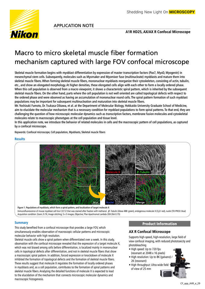
When this cell population is observed from a macro viewpoint, it shows a characteristic spiral pattern, which is inherited by the subsequent skeletal muscle fibers. On the other hand, parts where the cell population is not well oriented are called topological defects with respect to the ordered phase and were observed as having an accumulation of mononuclear round cells. The spiral pattern formation of such myoblast populations may be important for subsequent multinucleation and maturation into skeletal muscle fibers.
Mr. Yoshizuki Fumoto, Dr. Tsukasa Oikawa, et al. at the Department of Molecular Biology, Hokkaido University Graduate School of Medicine, aim to elucidate the molecular mechanism that is a necessary condition for myoblast populations to form spiral patterns. To that end, they are challenging the question of how microscopic molecular dynamics such as transcription factors, membrane fusion molecules and cytoskeletal molecules relate to macroscopic phenotypes at the cell population and tissue level.
In this application note, we introduce the behavior of related molecules in cells and the macroscopic pattern of cell populations, as captured by a confocal microscope.