尼康精机(上海)有限公司 | Chinese Mainland
- zh Change Region
- Global Site
卓越中心
The Nikon Center of Excellence at the Center for Gastrointestinal Biology (CGB) in Federal University of Minas Gerais (UFMG) is focused in providing imaging solutions to researchers that need to understand the mechanisms behind their projects imaging ions flux (i.e. calcium waves), immune cell migration and interaction and distribution and expression of several different molecules specially in their most native context: in vivo. For this, CGB has a very flexible imaging core that allows the imaging in vivo of single cells, organ slices and whole organs in a living mouse using multi-channel confocal intravital microscopy.
Nikon partnership has enhanced our ability to adapt stock microscopes to become high performance and accessible platforms for in vivo imaging. It is important to mention that conventional slices techniques, including 3D rendering of immunohistochemistry slices and basic fluorescence experiments can also be done using the same microscopes at our CofE. We make all efforts to serve the whole UFMG and other Minas Brazilian Universities, adapting our microscopes to the different demands.
The expertise, accessibility and advanced training provided by Nikon has allowed even very young students since the beginning of their graduating career to understand, learn and actually manipulate and use by themselves our Nikon confocal microscopes. Our imaging core has 2 confocals, and both that can be used for in vivo or ex vivo imaging. Using delicate surgical techniques and animal life support and care, we can image in high resolution, and even in three dimensions using automatic Z-stacks different cell behaviour in vivo during several hours in a living mouse. This produces amazing movies that have enhanced our knowledge in different areas.
email hidden; JavaScript is required
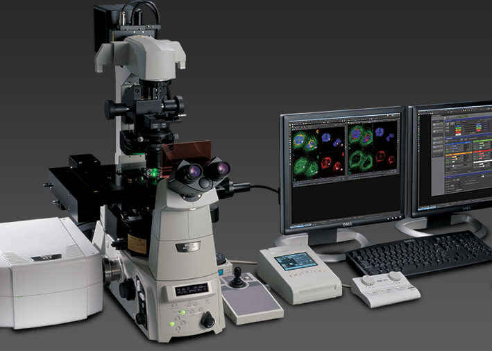
This high-speed A1Rsi resonant scanning confocal system features 32-channel spectral detector, optimized for live imaging of multiply labeled specimens.
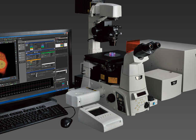
This C2 confocal is configured on a Ti inverted microscope and provides a fully integrated point scanning confocal well suited to many biomedical imaging applications.