


Centro de Excelência
The University of Western Australia
The Centre for Microscopy, Characterisation and Analysis (CMCA) is a core facility centre within The University of Western Australia, where research excellence is achieved by providing world-class science infrastructure and expertise to researchers and industry. Provision of such excellence is achieved through a focus on world-class facilities matched with expertise, and through a concept-to-publication User Pathway designed to meet the needs of researchers and industry through quality in training, acquisition and analysis.
Systems Available
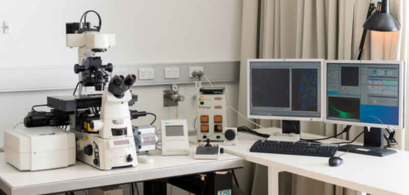
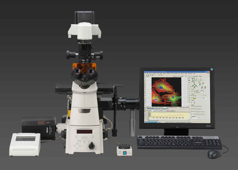
Nikon A1Si spectral detector confocal system plus TIRF on a Nikon Ti-E inverted motorised microscope
This inverted confocal microscope is equipped with 4 solid state lasers (405nm, 488nm, 561nm and 640nm), four photomultiplier fluorescence detectors, a transmission detector (can perform DIC, polarization or phase contrast imaging), as well as a 32-channel spectral detector. Imaging can be performed either in simultaneous or sequential mode, and we have a combination of high quality dry and immersion objective to suit your imaging needs. Complex imaging techniques are able to be performed on this system, including multiple fields, montaging, reflection and Fluorescent Recovery After Photo-bleaching (FRAP) microscopy.
Components
- Inverted Nikon TiE microscope
- Nikon PlanApo objectives 10x-40x, air and oil immersion
- Nikon PlanApo 60x WI IR, 1.27NA objective
- Nikon PlanApo 100x Oil immersion, 1.45NA objective
- Four 50mW solid state lasers – 405nm (violet), 488nm (blue), 561 (yellow), and 640nm (red)
- Two PMT detectors
- Two Gallium Arsenide Phosphatide (GaAsP) detectors
- Galvanometer and resonant scanners
- Three picosecond pulsed lasers – 405nm (violet), 485nm (blue) and 640nm (red)
- Picoquant Symphotime 64 analysis software
- Two PMA hybrid detectors
- Piezo stage
- Perfect Focus System 3
- NIS Elements software controlling confocal acquisition and lasers
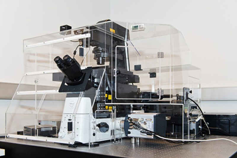
Nikon SIM (Ti2) (inverted)
Housed on a state-of-the-art Ti2 microscope, our SIM platform can acquire images with a resolution down to 100nm. Various objectives are available for this system, including 100x oil immersion with motorized correction collar, and 60x WI objective, specialized for live cell imaging. It is a four colour platform (405nm, 488nm, 561nm and 640nm solid state lasers), where images are acquired on an Andor iXon 897 EMCCD camera.
In addition to this, the system is capable of imaging in either 2D SIM, 3D SIM, 3D stack SIM or TIRF SIM mode for fixed or live cells (Tokai Hit stage top incubation system). Stability of the field of view over the imaging period is enabled by the Piezo stage and Perfect Focus System (PFS).
Components
- Inverted Nikon Ti2 microscope
- Nikon PlanApo objectives 10x-20x, air
- Nikon SR PlanApo IR 60x 1.27NA WI objective
- Nikon SR Apo TIRF 100x 1.49NA Oil immersion objective
- Four solid state lasers – 405nm (violet, 67mW), 488nm (blue, 120mW), 561 (yellow, 120mW), and 640nm (red, 80mW)
- Andor iXon897 EMCCD camera
- Three & four colour grating blocks for 60x WI & 100x Oil objectives
- Piezo stage
- NIS Elements software controlling confocal acquisition and lasers
- Tokai Hit stage-top incubation system
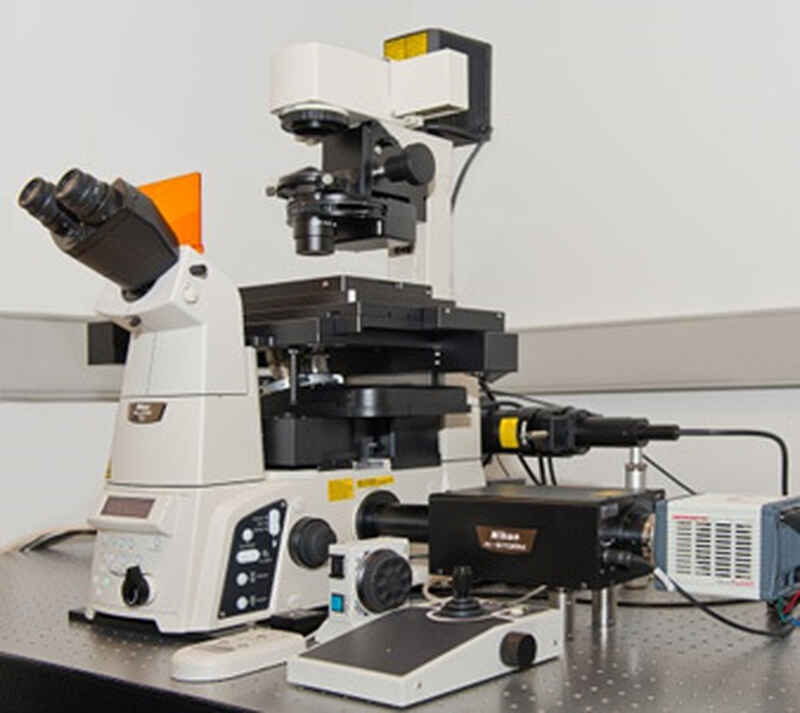
Nikon STORM (inverted)
Localisation imaging is the final super resolution microscope platform in our suite. Imaging with this modality exploits the photophysical behaviour of fluorophores to our N-STORM system is built on an inverted Ti-E microscope, fitted with four solid state lasers (405nm, 488nm, 561nm and 640nm) and a Hamamatsu OCRA FLASH 4 sCMOS camera, for high speed acquisition. C-STORM imaging can be performed on this platform, as well as the novel N-STORM imaging modality, where tandem dye pairs are used conjugated to samples utilising the high performance of Alexafluor 647 fluorescent dye.
The system is also equipped with Tokai Hit stage top incubator for live cell imaging, and Perfect Focus System (PFS) to prevent drift.
Components
- Inverted Nikon TiE microscope
- Nikon PlanApo objectives 10x-20x, air
- Nikon HP Apo TIRF 100x 1.49NA Oil immersion objective
- Four solid state lasers – 405nm (violet, 67mW), 488nm (blue, 120mW), 561 (yellow, 120mW), and 647nm (red, 300mW)
- Hamamatsu ORCA-Flash4.0 V3 sCMOS camera
- Piezo stage
- Perfect Focus System 3
- NIS Elements software controlling confocal acquisition and lasers
- Tokai Hit stage-top incubation system
Nikon Ti (inverted) with 6-colour fluorescence
This epifluorescence platform is utilising an inverted Ti-E microscope, with a special combination of filters and dichroics to be able to rapidly image six fluorescent probes. Using a Prior encoded motorised stage and an Andor iXon 885 EMCCD camera, whole section, six-coloured fluorescence imaging and analysis can be performed.