


Image courtesy of Laurence Pelletier Lab, LTRI
A1 HD25 / A1R HD25
Confocal Microscope System
Discontinued Replaced by AX / AX R with NSPARC
An Inside Look An Inside Look: How the A1 HD25/A1R HD25 achieved an industry-leading 25mm field of view
The A1 HD25/A1R HD25 confocal microscope has achieved the industry’s largest field of view, dramatically improving data throughput for confocal imaging. In this Q&A, we provide an inside look into the development and planning process behind this groundbreaking confocal.
How did this concept develop?
Nishida: In 2016, we achieved an unprecedented 25mm field of view with the Ti2 inverted microscope. Feedback from this product confirmed a high demand for large FOV cameras and detectors. In recent years, research has been trending towards examining biological processes within intact tissues, or even whole organisms, rather than in isolated cells. Accompanying this trend has been a rising need to be able to capture larger images without sacrificing resolution or detail. With this goal in mind, we designed a confocal system that would take full advantage of the Ti2’s 25mm FOV. Combined with its intrinsic optical sectioning capability, the new confocal design would enable users to not only capture larger images in XY but also to image thick samples.

Hiroshi Nishida
Joined Nikon in 1999. After working in international sales for 10 years, he worked at the company’s subsidiary in Europe for 6 years. Since 2015, he has been in charge of confocal microscopes in the Product Planning Department.
What are the advantages of the large field of view A1 HD25 and A1R HD25 confocal systems?
Nishida: The increased field of view allows the researcher to capture events occurring outside of the normal imaging area. For example, during time-lapse experiments carried out at high magnification, the researcher typically follows one to two cells over the course of the experiment. The enlarged field of view of the A1 HD25/A1R HD25 would enable researchers to capture the dynamics in neighboring cells that were previously obscured, while maintaining the same resolution and magnification. This ability may shed new light on the biological phenomena by providing larger contextual information. In addition, by collecting more data within each experiment, data fidelity or statistical robustness is improved. Image stitching can be used to expand the field of view but this method is slow and not conducive to capturing fast dynamics. It is important that a commercially available microscope can be used to capture large field-of-view images.

Masaru Horikoshi
Joined Nikon in 1989. He designed surveying instruments for 14 years. Since 2006, he has been designing confocal microscopes.
Horikoshi: Increasing the FOV from the traditional 18mm to 25mm results in approximately a two-fold increase in the amount of data captured in a single image. This is a significant increase in data throughput and would be an obvious advantage in high-content screening applications as well.

Achieves a field of view twice as large as conventional models
What were the challenges of developing this product?
Horikoshi: We had challenges when developing the scan lens. Simply increasing the diameter would affect image quality at the periphery of the field of view and introduce other aberrations. In order to expand the field of view and maintain our highest standards for image quality and field flatness, we had to re-design not only the optical system but also the glass formulation.
The development process required close collaboration with many departments including Manufacturing, Planning, and Development. One of Nikon's strengths is that we have an entirely integrated in-house production system for lenses, including glass making.
In addition to the scan lens, we made significant design changes to the entire optical system. Some of the challenges involved there included resolving errors induced by enlarging prisms and in increasing the speed of the galvanometer scanner to match its expanded control range.

Large diameter scan lens that expands the field of view (left)
Nishida: The development process was not simply about increasing the diameter. We wanted to maintain the high level of image quality known to be achieved by our A1 confocal series. Because the larger field of view requires stricter performance standards, we also had to fine-tune parts that did not enter the equation before.

We made full-scale improvements to achieve both a large field of view and high-quality images
What are the specific strengths of Nikon?
Nishida: We are committed to total optical performance, from raw glass to software. For example, one of Nikon's unique strengths is that we carefully examine not only the specifications of a standalone objective but also consider the overall optical performance when it is mounted on a microscope system.
Another example is Nikon's software, NIS-Elements. Our software not only controls Nikon equipment but is designed to easily control cameras and peripheral devices from other manufacturers, which allows users to completely customize and evolve their microscope system to match their application needs. This is a unique feature for a commercial software platform and a significant advantage for users.
Horikoshi: Our key strength is expandability. Nikon’s imaging devices are designed to be combined with one another to create unique imaging systems that address the custom needs of the researcher. For example, the A1 HD25/A1R HD25 confocal can be combined with a super-resolution system to enable users to easily capture large FOV images and super-resolve nanoscopic structures on the same microscope platform. Nikon’s systems enable users to seamlessly span nanoscopic to macroscopic observations.

How do you think the A1 HD25 and A1R HD25 will affect scientific research?
Horikoshi: I would be pleased if the large field of view of the A1 HD25 and A1R HD25 improves the efficiency of researchers’ experiments and helps them to clarify biological phenomena.
Nishida: I expect the A1 HD25 and A1R HD25 to be used for screening and drug efficacy testing because their larger field of view significantly increases the number of cells that can be captured and analyzed without sacrificing speed. I hope that the enhanced field of view will also provide new views into spatio-temporal dynamics of biological processes in live organisms and tissues.

Screening throughput is increased with a large field of view covering almost the entire area of a well
Recommended Model:
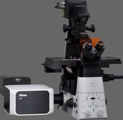
AX / AX R with NSPARC
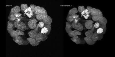
Nikon’s latest confocal microscope, leveraging deep learning-based AI tools to simplify acquisition and improve image quality. Fast scanning combined with a large 25 mm field of view enables high throughput imaging with unmatched edge-to-edge image quality. Super-resolution provided by the NSPARC detector.