
Paul Ronald Selvin, Ph.D.
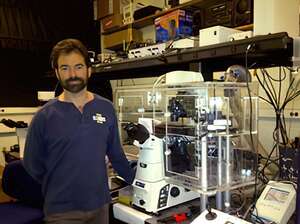
Professor, Department of Physics, University of Illinois at Urbana-Champaign
関連製品
- ECLIPSE Ti-E Inverted Microscope (現行機種:ECLIPSE Ti2)
Please tell us about your relationship with Nikon.
Nikon understands the uncertainty and sensitivity of my field.
I have worked closely with Nikon Instruments to build an imaging system that fully supports my research goals. For example, side by side with Nikon engineers and product managers, I tweaked the stage of the company’s Ti microscope so it moves in incredible plus/minus 10 nanometer increments—improving upon the former range of plus/minus 30 nanometers and also reducing vibrations. I also collaborated with the company to better control the temperature of the system (to prevent drift), using different lasers to enable viewing of different fluorophores, and viewing work with a very high numerical aperture objective.
I have come to depend on Nikon to work with me through many iterations of change to my systems to get them just right. The support I received from the company was nothing short of fantastic. If I had known then what I know now, every single one of my microscopes would be Nikon.
Please tell us about the development of FIONA.
It’s a partnership that really blossomed when I became the 2010 Nikon Fellow and worked closely with the company to develop a specific system for my work at the Marine Biological Laboratory. Nikon allowed me to take my custom microscope back to the lab and shortly thereafter we received a grant that allowed us to purchase it. Our team continue to innovate new ways to produce clear images of the tiny specimens we study. We invented FIONA, or Fluorescence Imaging with One Nanometer Accuracy. As the name implies, FIONA allows researchers to determine the position of a fluorophore to about a nanometer in the x-y plane, anywhere from a millisecond or longer, depending on its brightness.
FIONA coupled with creative dye selection and manipulation allows researchers to study cytoplasmic molecular motors, both in vitro and in vivo. Our team has captured the movements of these motors and found that they move by “walking,” one “foot” in front of the other with a stride ranging from 16 to 74 nanometers.
Please tell us about your other research.
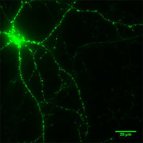
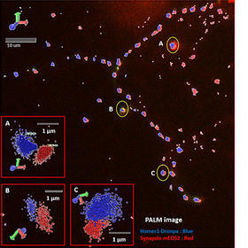
Our team has also developed a series of super—resolution techniques by separating the distance between dyes to achieve a better diffraction-limit of about 250 nm. By relying on successive photobleaching of the dyes—either permanently, or transiently—we have demonstrated that it is possible to get down to about 8 nm between two dyes, and approximately 50 nm measuring up to 20 dyes. We now use the system to study neuronal synapses and measure distances on DNA and RNA.
What do you think of Nikon and Nikon products?
When you’re in the business of single molecules, good accuracy and resolution is key. I count on our Nikon instrumentation to allow us to see cells in a consistent, reliable fashion. The customization, in which I collaborated with Nikon, has fine-tuned our equipment to create the ideal conditions for viewing these tiny samples. However, what I value most is the support I receive from Nikon. I have called up or e-mailed them so many times with various questions or challenges, and I always get a totally professional response in a rapid time-scale. That makes all the difference.